2. 中山大学第三附属医院,广东广州 510000;
3. 广西医科大学第二附属医院,广西南宁 530005;
4. 广西壮族自治区人民医院,广西南宁 530021
2. The Third Affiliated Hospital of Sun Yat-sen University, Guangzhou, Guangdong, 510000, China;
3. The Second Affiliated Hospital of Guangxi Medical University, Nanning, Guangxi, 530005, China;
4. Guangxi Zhuang Autonomous Region People′s Hospital, Nanning, Guangxi, 530021, China
目前,心血管疾病(Cardiovascular Diseases,CVD)是我国城乡居民疾病死亡的首要原因,其发病率及死亡率一直居高不下。2020年我国农村、城市居民死因中心血管疾病分别占48.00%和45.86%,即大约每5例死亡患者中就有2例是死于心血管疾病[1]。心血管影像对于心血管疾病诊治的重要性不言而喻,随着人工智能(Artificial Intelligence,AI)的快速发展,其在心血管领域,特别是在心血管影像分析中的应用展现出巨大的潜力。研究表明,利用机器学习等AI技术对心血管影像的图像采集、测量、报告及预测心血管疾病的预后赋能,其结果与领域内专家的分析结果基本一致,而且在分析速度等方面甚至能够超越专家[2-4]。尽管目前该领域研究数量不断增多,但鲜有学者进行系统的文献计量研究,而文献计量学可以在一定程度上系统评估某一领域、研究的发展过程及现状,并预测其发展趋势[5],故本文使用CiteSpace等文献计量学软件对AI应用于心血管影像的研究进行系统分析,旨在阐明该领域研究的基本情况及发展的趋势,为该领域学者提供参考。
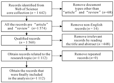
1 材料与方法 1.1 数据来源在2024年9月12日利用Web of Science (WOS)的核心合集数据库进行检索,查找并下载有关AI应用于心血管影像的研究文献,检索式如下:TS=(“imaging technique” OR “imaging techniques” OR “imaging”) AND (TS=(“cardiac” OR “intracardiac”)) AND (TS=“artificial intelligence” OR TS=“computational intelligence” OR TS=“deep learning” OR TS=“computer aided” OR TS=“machine learning” OR TS=“support vector machine” OR TS=“data learning” OR TS=“artificial neural network” OR TS=“digital image” OR TS=“convolutional neural network” OR TS=“evolutionary algorithms” OR TS=“feature learning” OR TS=“reinforcement learning” OR TS=“big data” OR TS=“image segmentation” OR TS=“hybrid intelligent system” OR TS=“recurrent neural network” OR TS=“natural language processing” OR TS=“bayesian network” OR TS=“bayesian learning” OR TS=“random forest” OR TS=“evolutionary algorithms” OR TS = “multiagent syste-m”),语言限制为“English”,文献类型限制为“article”和“review”,文献发表时间限制于2000年1月1日至2024年9月12日,共检索出1 560篇文献,为了确保数据准确性,2名研究者独立阅读文献的题目及摘要,对文献进行筛选,并交叉核对纳入的文献,当存在分歧时,由2名研究者通过讨论解决,对筛选出的文献进行查重后,最终纳入1 112篇文献,文献筛选流程见图 1。
|
| 图 1 文献筛选流程图 Fig. 1 Flow chart of literature screening |
1.2 统计学方法
使用Excel 2016绘制发文量的条形图并进行多项式拟合,使用CiteSpace 6.3.R1[6-7]、VOSviewer[8]、R语言的ggplot2对作者合作、机构合作、国家(地区合作)、关键词共现、文献共被引等进行分析并绘制图谱,以形象、直观地呈现出目前AI应用于心血管影像研究的整体情况、研究热点与发展趋势等,为今后的研究提供参考。
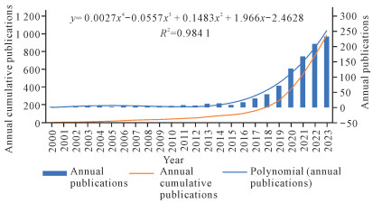
2 结果与分析 2.1 发文数量和趋势分析1 112篇文献中包括论著921篇(82.82%),综述191篇(17.18%)。2015年以前年发文量少且增长缓慢,年发文量最多的为2013年(13篇);2020年(127篇)为该领域研究首次突破100篇,自此该领域年发文量开始呈现大规模增长,2023年达232篇,约为2013年的18倍(图 2)。近4年发文量为总发文量的66.00%,多项式拟合结果显示,R2=0.984 1,表明发表年份与年发文量之间存在相关性。
|
| 图 2 年发文量变化趋势 Fig. 2 Trends of number of annual publications |
2.2 国家合作分析
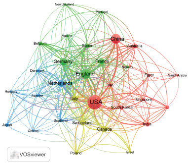
该领域发表的文献共来自70个国家(地区),发文量最多的国家(地区)为美国,发文量不少于30篇的国家(地区)有14个。发文量排在前10的国家(地区)的发文情况如表 1所示,发文量排在前5位的国家(地区)依次是美国、英国、中国、德国和荷兰,中国发文量位列第3(208篇);总被引用频次排在前5的国家(地区)依次是美国、英国、德国、中国和荷兰。从中介中心性来看,中国虽然发文量位居第3,但是中介中心性仅为0.11,位列第7。基于VOSviewer分析的国家(地区)间合作情况如图 3所示,图中节点之间的线条越粗表示两个节点间的合作关系越紧密,即总链接强度(Total Link Strength,TLS)越强[9],TLS排在前5的国家(地区)依次为美国、英国、荷兰、德国、意大利,中国排名第7。
| 排名Rank | 国家(地区)Country(region) | 发文量Number of publications | 占比/%Proportion/% | 总被引频次Total citations | 平均被引频次Average citations | 总链接强度TLS | 中介中心性Betweenness centrality | 首次出现时间First appearance time |
| 1 | USA | 441 | 30.58 | 10 290 | 23.33 | 499 | 0.28 | 2000 |
| 2 | UK | 224 | 15.53 | 6 364 | 28.41 | 345 | 0.22 | 2001 |
| 3 | China | 208 | 14.42 | 2 201 | 10.58 | 151 | 0.11 | 2006 |
| 4 | Germany | 120 | 8.32 | 3 064 | 25.53 | 207 | 0.13 | 2006 |
| 5 | Netherlands | 101 | 7.00 | 2 160 | 21.39 | 222 | 0.11 | 2004 |
| 6 | Canada | 93 | 6.45 | 1 631 | 17.54 | 139 | 0.15 | 2004 |
| 7 | Italy | 78 | 5.41 | 1 496 | 19.18 | 167 | 0.1 | 2000 |
| 8 | France | 72 | 4.99 | 1 162 | 16.14 | 115 | 0.19 | 2003 |
| 9 | Switzerland | 60 | 4.16 | 2 058 | 34.30 | 166 | 0.07 | 2004 |
| 10 | Spain | 45 | 3.12 | 1 200 | 26.67 | 96 | 0.03 | 2007 |
|
| 图 3 国家(地区)间的合作网络图谱 Fig. 3 Network of collaboration among countries(regions) |
2.3 机构合作分析
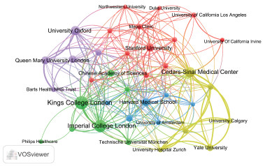
研究将AI应用于心血管影像的机构总共有1 905家,发文量排在前10的机构如表 2所示,这10家机构中,90%的机构来自英、美两国,中国科学院是发文量排名前10机构中唯一的中国机构。VOSviewer软件绘制的机构间合作网络图谱(图 4)表明,TLS排在前5的机构为伦敦国王学院、伦敦帝国理工学院、雪松西奈医学中心、牛津大学和斯坦福大学。中国对AI应用于心血管影像的研究发文量不少于11篇的机构如表 3所示,其中发文量超过20篇的机构仅有中国科学院。
| 排名Rank | 机构Institution | 发文量Number of publications | 总被引频次Total citations | 平均被引频次Average citations | 总链接强度TLS |
| 1 | King′s College London | 61 | 2 128 | 34.89 | 92 |
| 2 | Imperial College London | 54 | 3 439 | 63.69 | 97 |
| 3 | Cedars-Sinai Medical Center | 44 | 1 513 | 34.39 | 113 |
| 4 | University of Oxford | 36 | 1 590 | 44.17 | 66 |
| 5 | Stanford University | 31 | 1 097 | 35.39 | 50 |
| 6 | Harvard Medical School | 29 | 733 | 25.28 | 58 |
| 7 | Queen Mary University London | 29 | 1 026 | 35.38 | 81 |
| 8 | University College London | 27 | 593 | 21.96 | 48 |
| 9 | Yale University | 25 | 444 | 17.76 | 55 |
| 10 | Chinese Academy of Sciences | 24 | 622 | 25.92 | 41 |
|
| 图 4 机构间的合作网络图谱 Fig. 4 Network of collaboration among institutions |
| 排名Rank | 机构Institution | 发文量Number of publications | 总被引频次Total citations | 平均被引频次Average citations | 总链接强度TLS |
| 10 | Chinese Academy of Sciences | 24 | 622 | 25.92 | 41 |
| 16 | Shanghai Jiao Tong University | 19 | 356 | 18.74 | 29 |
| 45 | Fudan University | 11 | 418 | 38.00 | 24 |
| 46 | Huazhong University of Science and Technology | 11 | 89 | 8.09 | 13 |
| 49 | Sichuan University | 11 | 35 | 3.18 | 12 |
2.4 作者合作及共被引分析
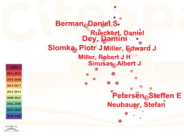
该领域研究涉及6 121名作者和22 203名共被引作者。发文量前10的作者如表 4所示。Damini Dey、Slomka Piotr J.等是该领域的主要作者。作者合作网络如图 5所示,其中Damini Dey、Berman Daniel S.、Slomka Piotr J.有较多合作,组成了一个AI结合心外膜脂肪组织及心肌灌注成像的研究网络[3, 10-12],而Neubauer Stefan、Petersen Steffen E.等组成的网络则将重点放在了研究心血管磁共振放射组学[13-17]上。
| 排名Rank | 作者Author | 发文量Number of publications | 被引频次Citations | 总链接强度TLS | 中介中心性Betweenness centrality |
| 1 | Damini Dey | 31 | 1 039 | 190 | 0.08 |
| 2 | Slomka Piotr J. | 27 | 1 232 | 172 | 0.02 |
| 3 | Berman Daniel S. | 24 | 908 | 179 | 0.08 |
| 4 | Petersen Steffen E. | 24 | 944 | 47 | 0.03 |
| 5 | Neubauer Stefan | 20 | 896 | 38 | 0.02 |
| 6 | Rueckert Daniel | 20 | 2 416 | 31 | 0.09 |
| 7 | Mmiller Edward J. | 19 | 290 | 165 | 0.02 |
| 8 | Sinusas Albert J. | 19 | 288 | 153 | 0.02 |
| 9 | Miller Robert J.H. | 17 | 280 | 135 | 0.02 |
| 10 | Liang Joanna X. | 13 | 206 | 135 | 0.00 |
|
| 图 5 作者合作网络 Fig. 5 Network of collaboration among authors |
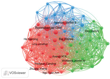
作者共被引网络如图 6所示。表 5显示Ronneberger Olaf共被引频次最多,排在第1,也是所有共被引作者中唯一共被引频次超过200次的作者,但中介中心性仅位列第4,Wolterink Jelmer M.共被引次数虽仅为第7位,但其中介中心性是所有作者中最高的。
|
| 图 6 作者共被引网络 Fig. 6 Network of co-citations among authors |
| 排名Rank | 作者Author | 共被引频次Co-citations | 总链接强度TLS | 中介中心性Betweenness Centrality |
| 1 | Ronneberger Olaf | 296 | 1 865 | 0.23 |
| 2 | Bai Wenjia | 174 | 1 570 | 0.06 |
| 3 | Bernard Olivier | 165 | 1 575 | 0.11 |
| 4 | Chen Chen | 142 | 1 120 | 0.00 |
| 5 | Petitjean Caroline | 126 | 1 195 | 0.66 |
| 6 | Petersen Steffen E. | 116 | 698 | 0.03 |
| 7 | Wolterink Jelmer M. | 115 | 1 603 | 0.85 |
| 8 | Zhuang Xiahai | 114 | 980 | 0.25 |
| 9 | Damini Dey | 111 | 994 | 0.05 |
| 10 | He Kaiming | 111 | 736 | 0.06 |
2.5 引用参考文献分析
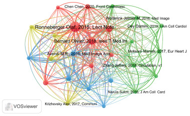
该领域研究涉及33 768篇参考文献,表 6展示了共被引排名前10文献的基本情况,其中共被引频次超过100的文献有3篇,其发表时间为2015-2018年,且研究主题均为将AI应用于心脏图像分割。图 7显示Ronneberger Olaf于2015年所著文章位于共被引网络中心。表 7为突现分析排名靠前的文献,该领域大量应用文献始于2012年。Ronneberger Olaf于2015所著文章“U-Net: convolutional networks for biomedical image segmentation”共被引频次及突显度排名第1,为该领域经典参考文献,该篇文章提供了一种用于自动图像分割的全卷积神经网络架构U-Net,U-Net可以在使用小样本量训练集的情况下较以往的模型更精确地分割图像[18]。
| 排名Rank | 文献Reference | 期刊/会议Journal/Conference | 第一作者First author | 发表年份Year | 共被引频次Co-citations | 总链接强度TLS |
| 1 | U-Net: convolutional networks for biomedical image segmentation | Lecture Notes in Computer Science | Ronneberger Olaf | 2015 | 249 | 723 |
| 2 | Deep learning techniques for automatic MRI cardiac multi-structures segmentation and diagnosis: is the problem solved? | IEEE Transactions on Medical Imaging | Bernard Olivier | 2018 | 148 | 613 |
| 3 | Automated cardiovascular magnetic resonance image analysis with fully convolutional networks | Journal of Cardiovascular Magnetic Resonance | Bai Wenjia | 2018 | 113 | 417 |
| 4 | Adam: a method for stochastic optimization | International Conference on Learning Representations | Kingma Diederik P. | 2015 | 82 | 223 |
| 5 | A review of segmentation methods in short axis cardiac MR images | Medical Image Analysis | Petitjean Caroline | 2011 | 82 | 289 |
| 6 | A combined deep-learning and deformable-model approach to fully automatic segmentation of the left ventricle in cardiac MRI | Medical Image Analysis | Avendi M.R. | 2016 | 79 | 398 |
| 7 | Deep learning for cardiac image segmentation: a review | Frontiers in Cardiovascular Medicine | Chen Chen | 2020 | 75 | 263 |
| 8 | Fully automated echocardiogram interpretation in clinical practice | Circulation | Zhang Jeffrey | 2018 | 68 | 246 |
| 9 | Deep residual learning for image recognition | 2016 IEEE Conference on Computer Vision and Pattern Recognition | He Kaiming | 2016 | 67 | 197 |
| 10 | Machine learning for prediction of all-cause mortality in patients with suspected coronary artery disease: a 5-year multicentre prospective registry analysis | European Heart Journal | Motwani Manish | 2017 | 63 | 207 |
|
| 图 7 文献共被引网络 Fig. 7 Network of co-citations among references |
| 排名Rank | 文献Reference | 期刊/会议Journal/Conference | 第一作者First author | 发表年份Year | 强度Strength | 开始时间Begin time | 结束时间End time |
| 1 | A review of segmentation methods in short axis cardiac MR images | Medical Image Analysis | Petitjean Caroline | 2011 | 8.13 | 2012 | 2017 |
| 2 | A combined deep-learning and deformable-model approach to fully automatic segmentation of the left ventricle in cardiac MRI | Medical Image Analysis | Avendi M.R. | 2016 | 11.74 | 2016 | 2020 |
| 3 | Right ventricle segmentation from cardiac MRI: a collation study | Medical Image Analysis | Petitjean Caroline | 2015 | 7.56 | 2015 | 2020 |
| 4 | U-Net: convolutional networks for biomedical image segmentation | Lecture Notes in Computer Science | Ronneberger Olaf | 2015 | 26.76 | 2018 | 2020 |
| 5 | Adam: a method for stochastic optimization | International Conference on Learning Representations | Kingma Diederik P. | 2015 | 8.75 | 2018 | 2020 |
| 6 | Machine-learning algorithms to automate morphological and functional assessments in 2D echocardiography | Journal of the American College of Cardiology | Narula Sukrit | 2016 | 7.95 | 2018 | 2020 |
| 7 | Deep learning | Nature | LeCun Yann | 2015 | 6.75 | 2018 | 2020 |
| 8 | Fully convolutional networks for semantic segmentation | 2015 IEEE Conference on Computer Vision and Pattern Recognition | Long Jonathan | 2015 | 5.95 | 2018 | 2020 |
| 9 | Combining deep learning and level set for the automated segmentation of the left ventricle of the heart from cardiac cine magnetic resonance | Medical Image Analysis | Ngo Tuan Anh | 2017 | 5.86 | 2018 | 2020 |
| 10 | CINENet: deep learning-based 3D cardiac CINE MRI reconstruction with multi-coil complex-valued 4D spatio-temporal convolutions | Scientific Reports | Küstner Thomas | 2020 | 7.15 | 2021 | 2024 |
2.6 期刊发文量及引用期刊情况
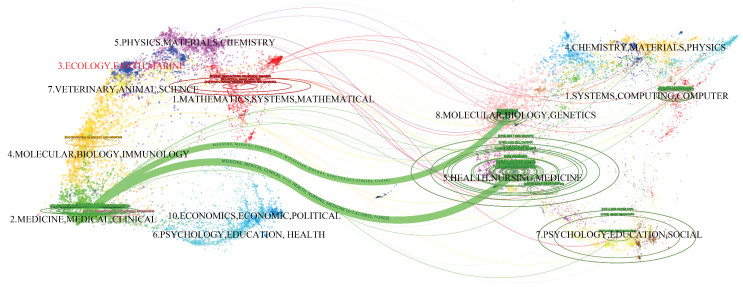
表 8显示的是发文量排名前10的期刊,其中Frontiers in Cardiovascular Medicine发文量最多,IEEE Transactions on Medical Imaging总被引频次最多,JACC-Cardiovascular Imaging影响因子最高。表 9列举了共被引频次排名前10的期刊,IEEE Transactions on Medical Imaging排名第1,影响因子最高的是European Heart Journal。期刊双叠加分析(图 8)结果表明,该领域主要引用路径有2条,分别为施引为“Medicine,Medical,Clinical”、被引为“Molecular,Biology,Genetics”以及施引为“Medicine,Medical,Clinical”、被引为“Health,Nursing,Medicine”;施引期刊的所属领域集中在“Medicine,Medical,Clinical”(“药物,医学,临床”)领域;被引期刊的所属领域集中在“Molecular,Biology,Genetics”(“分子,生物学,遗传学”)和“Health,Nursing,Medicine”(“健康,护理,药物”)领域。
| 排名Rank | 期刊名称Journal | 发文量Number of publications | 总被引频次Total citations | 国家Country | 影响因子(2022年)Impact factor(2022) | JCR分区(2022年)Journal citation reports partition(2022) | 总链接强度TLS |
| 1 | Frontiers in Cardiovascular Medicine | 73 | 1 014 | Switzerland | 3.6 | Q2 | 334 |
| 2 | IEEE Transactions on Medical Imaging | 47 | 2 119 | USA | 10.6 | Q1 | 249 |
| 3 | Medical Image Analysis | 30 | 1 263 | Netherlands | 10.9 | Q1 | 195 |
| 4 | IEEE Access | 26 | 340 | USA | 3.9 | Q2 | 80 |
| 5 | Medical Physics | 25 | 245 | USA | 3.8 | Q2 | 24 |
| 6 | Scientific Reports | 22 | 204 | UK | 3.8 | Q1 | 77 |
| 7 | Computer Methods and Programs in Biomedicine | 21 | 197 | Netherlands | 6.1 | Q1 | 66 |
| 8 | Computers in Biology and Medicine | 20 | 199 | USA | 7.7 | Q1 | 109 |
| 9 | European Radiology | 20 | 277 | Germany | 5.9 | Q1 | 50 |
| 10 | JACC-Cardiovascular Imaging | 20 | 1 083 | USA | 14 | Q1 | 103 |
| 排名Rank | 期刊Journal | 共被引频次Co-citations | 国家Country | 影响因子(2022年)Impact factor(2022) | JCR分区(2022年)Journal citation reports partition(2022) | 总链接强度TLS |
| 1 | IEEE Transactions on Medical Imaging | 2 205 | USA | 10.6 | Q1 | 110 703 |
| 2 | Journal of the American College of Cardiology | 1 790 | USA | 24.4 | Q2 | 98 314 |
| 3 | Lecture Notes in Computer Science | 1 782 | USA | — | — | 89 915 |
| 4 | Medical Image Analysis | 1 673 | Netherlands | 10.9 | Q1 | 85 818 |
| 5 | Circulation | 1 519 | USA | 37.8 | Q1 | 82 693 |
| 6 | JACC-Cardiovascular Imaging | 1 427 | USA | 14.0 | Q1 | 82 816 |
| 7 | Journal of Cardiovascular Magnetic Resonance | 1 347 | USA | 6.4 | Q1 | 75 978 |
| 8 | Magnetic Resonance in Medicine | 1 336 | USA | 3.3 | Q2 | 76 293 |
| 9 | European Heart Journal | 936 | UK | 39.3 | Q1 | 53 096 |
| 10 | Radiology | 895 | USA | 19.7 | Q1 | 54 384 |
|
| 图 8 期刊双叠加图谱 Fig. 8 A dual-map overlap of journals |
2.7 关键词分析
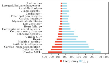
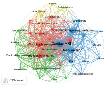
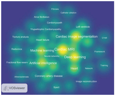
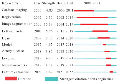
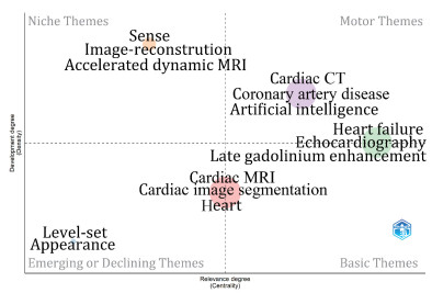
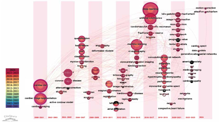
本文共纳入3 717个关键词,图 9总结了出现频次最高的前20个关键词。其中,“心血管磁共振”“深度学习”“心血管图像分割”“人工智能”“机器学习”是出现频次最高的5个关键词,这与本文研究主题一致。在图 10中,关键词的大小代表了其出现频次,“机器学习”和“深度学习”处于网络的中心位置。同时,图 11也表明,“机器学习”“深度学习”“心脏”“心血管磁共振”和“心血管图像分割”等关键词区域具有较高的热度。图 10和图 11表明,“机器学习”和“深度学习”是当前该领域的核心研究方法,而将其应用于心血管磁共振影像分割则是一个重要的研究热点。如图 12所示,该领域关键词主要分为13个聚类,而这13个聚类可进一步归属于三大研究领域——人工智能技术(#2深度学习、#8卷积神经网络、#9人工智能、#10机器学习)、心脏影像工具(#0图像重建、#4CT、#6心血管磁共振、#7图像分割、#11三维左心室重建、#13三维超声心动图)和心脏疾病(#1心肌梗死、#3冠状动脉疾病)。突现度排名前10的关键词如图 13所示,“图像分割”的出现始于2000年,出现频次于2004年突然增高,并持续至2019年,其突现强度也是最高的,这表明图像分割是研究人员关注的重点;“神经网络”“模型”“动脉疾病”“特征提取”为2016年以来的主要突现词,表明这些研究课题近年来受到了广泛关注。图 14显示,该领域主题共有4个,其中Cardiac CT、Coronary artery disease、Artificial intelligence为该领域的核心主题;Sense、Image-reconstruction、Accelerated dynamic MRI为该领域的边缘主题;Cardiac MRI、Cardiac image segmentation、Heart为该领域的新兴&基本主题,且对该领域有一定的重要性;Heart failure、Echocardiography、Late gadolinium enhancement为该领域的核心&基本主题,可能是将来该领域重要的主题。在图 15中,心血管磁共振及心血管影像分割、心血管电子计算机断层扫描(Computed Tomography,CT)、机器学习、深度学习、人工智能卷积神经网络是该领域重要的节点。根据节点的分布情况,可以将该领域研究以2014-2015年为界划分为两个主要阶段:在第一阶段(2000-2015年),该领域研究重点是基于心血管CT及磁共振成像(Magnetic Resonance Imaging,MRI)的图像分割、冠脉病变及心力衰竭,但受限于科技水平,该阶段研究人员使用的分析技术有限;而在第二阶段(2015年至今),由于科技的发展,该领域研究得到了AI的加持,深度学习因其卓越的性能成为这一阶段关键词时区图内出现频次最多的节点。
|
| 图 9 出现频次排名前20关键词 Fig. 9 Top 20 keywords with the highest frequency |
|
| 图 10 关键词共现图谱 Fig. 10 Map of keyword co-occurrence |
|
| 图 11 关键词密度图谱 Fig. 11 Map of keyword density |

|
| 图 12 关键词聚类图谱 Fig. 12 Map of keyword clustes |
|
| 图 13 突现度排名前10关键词 Fig. 13 Top 10 keywords with the strongest citation bursts |
|
| 图 14 主题地图 Fig. 14 Thematic map |
|
| 图 15 关键词时区图 Fig. 15 Map of keyword time zone |
3 讨论
近年来发展迅猛的AI技术在医学影像领域的优势不断被释放,正逐步引领医学影像技术走向前所未有的大变革,将AI应用于心血管影像的研究不断增加。本文利用文献计量学的相关技术,使用VOSviewer、CiteSpace、R语言等软件首次梳理并归纳AI在心血管影像领域的应用和发展,为领域研究人员展示其发展历史,并预测新的研究热点。
3.1 提升学术影响力,整合、扩展区域研究力量发文量分析和国家、机构共现分析结果表明,由于AI技术及心血管成像精密度的不断提高,心血管疾病的图像获取、解释以及决策的成本降低[3],因此心血管影像的相关AI技术还将在美、英等高学术影响力国家研究人员的主导下不断向前推进,他们未来的发文量还将继续增加,国内虽然在该领域有一定的学术影响力,但是未来应该在兼顾发文数量的同时进一步提高研究质量及学术影响力。目前国内对该领域的研究存在较大的地域差异,学术影响力较大的机构主要位于北京、上海等经济发达城市,并且国内各机构之间缺乏合作。因此我国若想在未来提高在该领域的研究质量及学术影响力,首先,需要加强与影响力较大国家、机构的学术合作;其次,需要国内影响力较大的机构分享经验,实现技术的传—带—帮,并加强区域内及区域间合作;最后,还需要增加对经济落后地区相关科研技术及资金的投资,以期这些机构将来有可能成为这一领域的重要参与者。
3.2 图像分割仍是当前热点文献共被引分析可以在一定程度上反映该领域研究热点。该领域共被引频次最高的文献“U-Net: convolutional networks for biomedical image segmentation”中提出一种适配医学图像分割的架构,并且此后其作为基础架构不断被完善[19-22];2021年发表在Nature Methods的文献“nnU-Net: a self-configuring method for deep learning-based biomedical image segmentation”中所提出的架构就是基于U-Net架构而设计的一个自适应架构,能够自动根据处理对象进行自我配置以完成任务[23]。这在一定程度上代表了目前心血管影像AI模型的发展趋势,即该领域模型必须能够在小数据集上进行训练。
3.3 AI赋能无创影像检查通过对热门研究主题的识别,关键词共现分析可以帮助研究者更好地了解研究热点[24]。与临床医师相比,心血管影像相关的AI算法不会受限于临床经验,并且处理图像的速度大大加快。不同的影像方法与AI的结合为心血管疾病的管理提供了新的可能性。
3.3.1 AI应用于超声心动图超声心动图因其非侵入性、简便经济的特点被广泛应用于心血管疾病的影像检测,但是由于其图像质量易受噪声干扰,因此之前在心血管疾病的管理中作用有限。然而由于AI技术的应用,目前超声心动图对心脏进行分割的准确率已经超过90%。在对心脏进行三维测量时,AI模型能够同时捕获面积变化分数、三尖瓣环平面收缩期离散度等多个二维超声心动图参数。AI加持的三维超声心动图克服了三维超声的操作复杂性以及对手动输入的依赖性,并且进一步完善了心内膜检测或系数构建[25]。He等[26]发现使用AI评估左心室射血分数与人工评估的准确性相当,并且评估所需时间更短,可应用于心力衰竭的诊断和管理。另外,超声心动图可通过评估心壁运动检测冠脉病变及心肌梗死[27-28],Guo等[29]提出了一个用于检测心肌功能和左室的机器学习模型,用以检测冠脉病变,敏感性达到0.95,尽管由于其特异性过低而易导致假阳性,但仍为后续的研究提供了新的方向。除了在临床方面的应用,未经训练的护士在AI辅助下使用Point-of-care ultrasound可以准确测量心室大小、功能和心包积液[25]。
3.3.2 AI应用于CT在心血管CT领域,AI展现出巨大潜力。借助神经网络技术,AI在冠状动脉钙化评分和心外膜脂肪组织测量方面的表现已可媲美专家手动测量,且耗时显著缩短,有助于冠心病人群的筛查[30-32]。目前,国内冠脉狭窄评估仍以有创冠脉造影(Invasive Coronary Angiography,ICA)为主,但基于CT冠脉血管成像的血流储备分数(CT derived Fractional Flow Reserve,CT-FFR)因其具有无创性、计算时间短、安全性较高且费用较低的优势而受到关注。2018年已有指南建议CT-FFR可作为是否进行介入治疗的依据[33]。研究表明,基于机器学习的CT-FFR优化了患者治疗决策,降低了中度冠脉狭窄(25%-80%狭窄)患者的ICA转诊率及ICA中非阻塞性病变的发生率,并表现出良好的预后效果[34]。
3.3.3 AI应用于MRI心血管磁共振(Cardiovascular Magnetic Resonance,CMR)在心脏疾病诊治中的优势显著,但其检查过程耗时较长,需构建复杂序列、配合患者呼吸周期,且晚期钆增强成像需在特定时间点进行。研究表明,结合深度学习的“一体化/实时心血管磁共振”可有效解决这些问题。例如,通过生成磁共振指纹识别(Magnetic Resonance Fingerprinting,MRF)词典,所需时间从158.0 s缩短至0.8 s,未来甚至可能在亚秒级内获取更多组织特性,助力临床转化[35-36]。此外,CMR可无创评估心肌灌注,有望成为冠脉疾病管理的重要技术。目前已有研究开发出基于图像配准和分割的算法,实现CMR心肌灌注成像的自动评估,其定量灌注值与手动评估结果高度一致,且显著减少用户交互时间[37]。
尽管有创检测仍是心血管疾病评估的“金标准”,但随着深度学习等AI算法的引入,超声、CT和CMR等技术在心血管领域的潜力显著增强。AI的结合大幅提升了这些影像工具的无创评估能力,增强了其在心血管疾病管理中的作用。未来研究重点或将集中于进一步探索AI与各类心血管影像技术的深度融合,以持续提升无创评估的准确性和效率。
4 结论本文分析结果表明,AI在心血管影像中的应用显著提升了诊断的准确性和效率,尤其是在超声心动图、CT和MRI等技术中。通过AI算法的引入,医生能够更快速、精确地识别和评估心血管疾病,从而改善患者的治疗决策。随着AI技术的不断进步和数据的积累,在“健康中国”及“一带一路”的背景下,如何进一步通过结合AI以提高心血管影像技术对心血管疾病识别及评估的准确性、便捷性和效率是未来的研究重点,研究人员应聚焦于这些问题,致力于提高区域合作,特别关注心血管图像分割及冠脉疾病的图像诊断、治疗等方向,以进一步提高我国心血管医学研究实力,并为心血管疾病的管理开辟新的方向。但是,本文仅纳入WOS数据库中的英文文献,可能遗漏其他来源的重要文献,同时,由于数据库持续更新,近期发表的高质量文献可能因引用频次较低而未能充分体现其价值。
| [1] |
中国心血管健康与疾病报告编写组. 中国心血管健康与疾病报告2022概要[J]. 中国循环杂志, 2023, 38(6): 583-612. DOI:10.3969/j.issn.1000-3614.2023.06.001 |
| [2] |
王家鑫, 赵世华. 人工智能在心血管疾病影像学领域中的应用[J]. 中华心血管病杂志, 2021, 49(11): 1063-1068. DOI:10.3760/cma.j.cn112148-20210730-00639 |
| [3] |
DEY D, SLOMKA P J, LEESON P, et al. Artificial intelligence in cardiovascular imaging[J]. Journal of the American College of Cardiology, 2019, 73(11): 1317-1335. DOI:10.1016/j.jacc.2018.12.054 |
| [4] |
DAWES T J W, DE MARVAO A, SHI W Z, et al. Machine learning of three-dimensional right ventricular motion enables outcome prediction in pulmonary hypertension: a cardiac MR imaging study[J]. Radiology, 2017, 283(2): 381-390. DOI:10.1148/radiol.2016161315 |
| [5] |
ZHAO J, YU G Y, CAI M X, et al. Bibliometric analysis of global scientific activity on umbilical cord mesenchymal stem cells: a swiftly expanding and shifting focus[J]. Stem Cell Research & Therapy, 2018, 9(1): 32. |
| [6] |
SYNNESTVEDT M B, CHEN C M, HOLMES J H. CiteSpace Ⅱ: visualization and knowledge discovery in bibliographic databases[J]. AMIA Annual Symposium Proceedings AMIA Symposium, 2005, 2005: 724-728. |
| [7] |
CHEN C M, DUBIN R, KIM M C. Emerging trends and new developments in regenerative medicine: a scientometric update (2000-2014)[J]. Expert Opinion on Biological Therapy, 2014, 14(9): 1295-1317. DOI:10.1517/14712598.2014.920813 |
| [8] |
VAN ECK N J, WALTMAN L. Software survey: VOSviewer, a computer program for bibliometric mapping[J]. Scientometrics, 2010, 84(2): 523-538. DOI:10.1007/s11192-009-0146-3 |
| [9] |
董娜, 崔婷, 王露露, 等. 人工智能在胃癌诊治中的研究趋势: 20年的文献计量学分析[J]. 中国全科医学, 2024, 27(4): 493-501. DOI:10.12114/j.issn.1007-9572.2022.0902 |
| [10] |
EISENBERG E, MCELHINNEY P A, COMMAN- DEUR F, et al. Deep learning-based quantification of epicardial adipose tissue volume and attenuation predicts major adverse cardiovascular events in asymptomatic subjects[J]. Circulation: Cardiovascular Imaging, 2020, 13(2): e009829. DOI:10.1161/CIRCIMAGING.119.009829 |
| [11] |
COMMANDEUR F, SLOMKA P J, GOELLER M, et al. Machine learning to predict the long-term risk of myocardial infarction and cardiac death based on clinical risk, coronary calcium, and epicardial adipose tissue: a prospective study[J]. Cardiovascular Research, 2020, 116(14): 2216-2225. DOI:10.1093/cvr/cvz321 |
| [12] |
RIOS R, MILLER R J H, HU L H, et al. Determining a minimum set of variables for machine learning cardiovascular event prediction: results from REFINE SPECT registry[J]. Cardiovascular Research, 2022, 118(9): 2152-2164. DOI:10.1093/cvr/cvab236 |
| [13] |
CAMPELLO V M, GKONTRA P, IZQUIERDO C, et al. Multi-centre, multi-vendor and multi-disease cardiac segmentation: the M & Ms challenge[J]. IEEE Transactions on Medical Imaging, 2021, 40(12): 3543-3554. DOI:10.1109/TMI.2021.3090082 |
| [14] |
BAI W J, SINCLAIR M, TARRONI G, et al. Automated cardiovascular magnetic resonance image analysis with fully convolutional networks[J]. Journal of Cardiovascular Magnetic Resonance, 2018, 20(1): 65. DOI:10.1186/s12968-018-0471-x |
| [15] |
RAISI-ESTABRAGH Z, IZQUIERDO C, CAMPELLO V M, et al. Cardiac magnetic resonance radiomics: basic principles and clinical perspectives[J]. European Heart Journal-Cardiovascular Imaging, 2020, 21(4): 349-356. DOI:10.1093/ehjci/jeaa028 |
| [16] |
GUO F M, NG M, GOUBRAN M, et al. Improving cardiac MRI convolutional neural network segmentation on small training datasets and dataset shift: a continuous kernel cut approach[J]. Medical Image Analysis, 2020, 61: 101636. DOI:10.1016/j.media.2020.101636 |
| [17] |
XIA Y, RAVIKUMAR N, GREENWOOD J P, et al. Super-resolution of cardiac MR cine imaging using conditional GANs and unsupervised transfer learning[J]. Medical Image Analysis, 2021, 71: 102037. DOI:10.1016/j.media.2021.102037 |
| [18] |
RONNEBERGER O, FISCHER P, BROX T. U-Net: convolutional networks for biomedical image segmentation[C]//NAVAB N, HORNEGGER J, WELLS W M, et al. Medical image computing and computer-assisted intervention-MICCAI 2015: 18th international conference. Munich: Springer, 2015: 234-241.
|
| [19] |
LI Y Z, WANG Y, HUANG Y H, et al. RSU-Net: U-Net based on residual and self-attention mechanism in the segmentation of cardiac magnetic resonance images[J]. Computer Methods and Programs in Biomedicine, 2023, 231: 107437. DOI:10.1016/j.cmpb.2023.107437 |
| [20] |
YU X H, CHEN J X, FANG B, et al. Cardiac LGE MRI segmentation with cross-modality image augmentation and improved U-Net[J]. IEEE Journal of Biomedical and Health Informatics, 2023, 27(2): 588-597. DOI:10.1109/JBHI.2021.3139591 |
| [21] |
CUI H F, LI Y, JIANG L, et al. Improving myocardial pathology segmentation with U-Net++ and EfficientSeg from multi-sequence cardiac magnetic resonance images[J]. Computers in Biology and Medicine, 2022, 151: 106218. DOI:10.1016/j.compbiomed.2022.106218 |
| [22] |
CUI H F, CHANG Y W, JIANG L, et al. Multiscale attention guided U-Net architecture for cardiac segmentation in short-axis MRI images[J]. Computer Methods and Programs in Biomedicine, 2021, 206: 106142. DOI:10.1016/j.cmpb.2021.106142 |
| [23] |
ISENSEE F, JAEGER P F, KOHL S A A, et al. nnU-Net: a self-configuring method for deep learning-based biomedical image segmentation[J]. Nature Methods, 2021, 18(2): 203-211. DOI:10.1038/s41592-020-01008-z |
| [24] |
SHEN Z F, HU J T, WU H Y, et al. Global research trends and foci of artificial intelligence-based tumor pathology: a scientometric study[J]. Journal of Translational Medicine, 2022, 20(1): 409. DOI:10.1186/s12967-022-03615-0 |
| [25] |
QIN Y H, QIN X H, ZHANG J, et al. Artificial intelligence: the future for multimodality imaging of right ventricle[J]. International Journal of Cardiology, 2024, 404: 131970. DOI:10.1016/j.ijcard.2024.131970 |
| [26] |
HE B, KWAN A C, CHO J H, et al. Blinded, randomized trial of sonographer versus AI cardiac function assessment[J]. Nature, 2023, 616(7957): 520-524. DOI:10.1038/s41586-023-05947-3 |
| [27] |
MURAKI R, TERAMOTO A, SUGIMOTO K, et al. Automated detection scheme for acute myocardial infarction using convolutional neural network and long short-term memory[J]. PLoS One, 2022, 17(2): e0264002. DOI:10.1371/journal.pone.0264002 |
| [28] |
MEDVEDOFSKY D, MOR-AVI V, AMZULESCU M, et al. Three-dimensional echocardiographic quantification of the left-heart chambers using an automated adaptive analytics algorithm: multicentre validation study[J]. European Heart Journal - Cardiovascular Imaging, 2018, 19(1): 47-58. DOI:10.1093/ehjci/jew328 |
| [29] |
GUO Y, XIA C X, ZHONG Y, et al. Machine learning-enhanced echocardiography for screening coronary artery disease[J]. BioMedical Engineering OnLine, 2023, 22(1): 44. DOI:10.1186/s12938-023-01106-x |
| [30] |
XU B, KOCYIGIT D, GRIMM R, et al. Applications of artificial intelligence in multimodality cardiovascular imaging: a state-of-the-art review[J]. Progress in Cardiovascular Diseases, 2020, 63(3): 367-376. DOI:10.1016/j.pcad.2020.03.003 |
| [31] |
LEHKER A, MUKHERJEE D. Coronary calcium risk score and cardiovascular risk[J]. Current Vascular Pharmacology, 2020, 19(3): 280-284. DOI:10.2174/1570161118666200403143518 |
| [32] |
李雁鸣, 沈成兴, 申锷. 心外膜脂肪组织与心血管疾病的研究进展[J]. 中华心血管病杂志, 2022, 50(7): 723-727. DOI:10.3760/cma.j.cn112148-20220527-00417 |
| [33] |
中华医学会放射学分会心胸学组, 中国医师协会放射医师分会心血管学组, 北京医学会放射学分会心血管学组. CT血流储备分数操作规范及临床应用中国专家共识[J]. 中华放射学杂志, 2023, 57(7): 711-722. DOI:10.3760/cma.j.cn112149-20221215-01005 |
| [34] |
QIAO H Y, TANG C X, SCHOEPF U J, et al. One- year outcomes of CCTA alone versus machine learning-based FFRCT for coronary artery disease: a single-center, prospective study[J]. European Radiology, 2022, 32(8): 5179-5188. DOI:10.1007/s00330-022-08604-x |
| [35] |
CHRISTODOULOU A G, CRUZ G, ARAMI A, et al. The future of cardiovascular magnetic resonance: all-in-one vs.real-time (Part 1)[J]. Journal of Cardiovascular Magnetic Resonance, 2024, 26(1): 100997. DOI:10.1016/j.jocmr.2024.100997 |
| [36] |
CONTIJOCH F, RASCHE V, SEIBERLICH N, et al. The future of CMR: all-in-one vs.real-time CMR (part 2)[J]. Journal of Cardiovascular Magnetic Resonance, 2024, 26(1): 100998. DOI:10.1016/j.jocmr.2024.100998 |
| [37] |
WENG A M, RITTER C O, LOTZ J, et al. Automatic postprocessing for the assessment of quantitative human myocardial perfusion using MRI[J]. European Radiology, 2010, 20(6): 1356-1365. DOI:10.1007/s00330-009-1684-z |


